CN
CN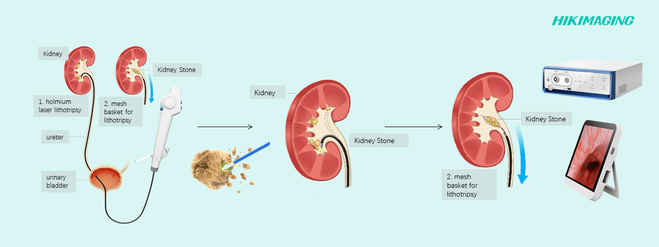
Hikimaging Ureteroscopy Solution——High-definition Vision Facilitates Ureteroscopic Lithotripsy
In the field of urology, ureteroscopy has become an important instrument for minimally invasive treatment of upper urinary tract stones. The flexible ureteroscope can directly explore the location, size, shape and surrounding mucosal condition of stones through the natural cavity of the human body (urethra-bladder-ureter). Compared with traditional percutaneous nephrolithotomy(pcnl), FURS significantly reduces the risk of bleeding and postoperative pain, shortens the recovery time, and has become the preferred treatment option for many patients with kidney stones. In FURS, clear and stable endoscopic imaging is the key to successful surgery. Blurred and noisy images will make it more difficult for doctors to identify stones and distinguish between tiny blood vessels and mucosal structures, which will not only prolong the operation time, but also pose the risk of accidental injury to tissues and residual stones. In this issue, we will use the actual intraoperative images of kidney stone holmium laser lithotripsy performed by “Hikimaging ureteroscopy solution”to intuitively experience the surgical process and the technological innovation brought about by high-definition imaging. Ureteroscopy to explore the renal pelvis and accurately locate stones The ureteroscope needs to be fully lubricated before insertion to reduce damage. Under the guidance of the guide wire, the target position is successfully reached through the urethra, bladder, and ureter. Doctors can flexibly control the endoscope through foot pedals or handles to systematically explore and locate the stones. https://www.hikimaging.com/wp-content/uploads/2025/07/1输尿管镜探查肾盂,找到结石.mp4 2. Ureteroscopy to explore the renal pelvis and accurately locate stones After confirming the target stone, the holmium laser fiber is inserted through the working channel of the soft mirror. The laser spot is precisely focused on the surface of the stone, and the “fragmentation” strategy is used to break the stone into sufficiently small particles. https://www.hikimaging.com/wp-content/uploads/2025/07/2剪辑版本.mp4 3. Postoperative reexamination to ensure that there are no “stones” After lithotripsy is completed, the renal pelvis and target calyces are examined again with a ureteroscope to ensure that there are no residues and to check the degree of tissue damage. https://www.hikimaging.com/wp-content/uploads/2025/07/3剪辑版本.mp4 Hikimaging ureteroscope solution, through the unique super-resolution technology, effectively improves the clarity of electronic mirror imaging. It can be used in combination with a variety of host options such as portable host and dual-mirror host, and can be flexibly used in consulting rooms and operating rooms. It provides a powerful tool for urologists and allows minimally invasive surgery to move further into a new era of “seeing clearly and doing accurately”. DownLoad
Hikimaging multi-color fluorescence for lymphadenectomy in gynecological cancer
Lymphadenectomy in gynecological cancer has long faced the challenge of accurate identification and complete removal. Micro-metastases, anatomical variations, and the hidden nature of deep lymph nodes make it very easy for traditional methods that rely on palpation and visual inspection to miss diseased tissues, affecting patient prognosis. Fluorescence imaging technology improves the accuracy and safety of lymphadenectomy through real-time visualization. Green fluorescence: real-time imaging, rapid targeting of target lymph nodes Doctors do not need to rely on preoperative imaging memory or repeated palpation. The fluorescent screen directly displays the location of the target lymph nodes. Especially for obese patients or superficial lymph nodes, the green fluorescence can quickly guide the surgeon to the target, shorten the operation time, and clearly mark the lymph node boundaries to reduce misoperation of surrounding normal blood vessels and nerves. https://www.hikimaging.com/wp-content/uploads/2025/07/1.绿色荧光:快速锁定淋巴结位置-2.mp4 White and multi-color fluorescence switching: Targeting deep lymph nodes Accurate identification of deep pelvic lymph nodes (such as obturator foramen and iliac vessels) is a difficult point in gynecological cancer surgery. Huiying’s white light and multi-color fluorescence switching function can clearly “light up” the previously hidden lymph nodes, avoiding missing key lesions. https://www.hikimaging.com/wp-content/uploads/2025/07/2.白光和多色荧光切换:锁定深处淋巴结.mp4 Multi-color fluorescence: Assists in complete removal of lymph nodes By observing the continuity and integrity of the fluorescent signal, the surgeon can intuitively determine whether the lymph node capsule is destroyed, whether satellite lesions exist, and whether the lymphatic drainage area has been cleared thoroughly enough to minimize the risk of residual tiny lesions. https://www.hikimaging.com/wp-content/uploads/2025/07/3.多色荧光:助力完整切除淋巴结-2.mp4 Hikimaging has launched a series of fluorescent products such as 4K fluorescent and 3D fluorescent, which greatly improve the development effect through algorithms. At the same time, the self-developed optical mount effectively suppresses stray light interference and significantly improves the problem of fluorescent light leakage. DownLoad

Hikimaging assists single incision laparoscopic cholecystectomy
Traditional laparoscopic surgery requires 3-4 incisions, which is very traumatic and slow to recover from. Single incision laparoscopic surgery (SILS) only ues a single incision to complete the surgery, which greatly reduces the trauma of surgery and greatly improves the patient’s recovery speed after surgery. This issue explains the entire SILS surgery process. Step 1: Dissect the gallbladder triangle: Accurately locate the gallbladder triangle area. Dissect the gallbladder triangle to expose clear blood vessels and bile duct structures. https://www.hikimaging.com/wp-content/uploads/2025/06/单孔腹腔镜胆囊切除术-解剖胆囊三角1.mp4 Step 2: Clamp and cut off the cystic artery:Use clamping instruments to block blood flow, avoid bleeding during surgery, and ensure a clear surgical field of view https://www.hikimaging.com/wp-content/uploads/2025/06/单孔腹腔镜胆囊切除术-夹闭胆囊动脉1.mp4 Step 3: Clamp and cut off the cystic duct: Safely separate and cut off the cystic duct to completely disconnect the lesion from the bile duct system. However, the integrity of the bile duct system must be ensured. https://www.hikimaging.com/wp-content/uploads/2025/06/单孔腹腔镜胆囊切除术-夹闭胆囊管.mp4https://www.hikimaging.com/wp-content/uploads/2025/06/单孔腹腔镜胆囊切除术-切断胆囊管.mp4 Step 4: Separate the gallbladder:Gently peel off the gallbladder bed and completely remove the diseased gallbladder https://www.hikimaging.com/wp-content/uploads/2025/06/单孔腹腔镜胆囊切除术-分离胆囊1.mp4 Hikimaging endoscope system empowers surgeons with 4K imaging system + intelligent equipment, making cholecystectomy safer, more precise and more efficient ! DownLoad

VF Technology
Background: Focal length is closely related to depth of field. As a key factor affecting the range of image clarity, precise focal length adjustment can enable the target object to be clearly imaged on the image sensor.Depth of field refers to the object distance range corresponding to the clear imaging on a fixed image plane. Due to factors such as the improvement of the imaging system’s resolution and the increase of the magnification, the depth of field range will be limited. The limitation of the depth of field makes it impossible for traditional imaging systems to achieve clear imaging of objects with large depth differences under fixed focus. In the use of medical endoscopes, it is often necessary to change the imaging magnification flexibly. Low magnification is used to observe images within a larger range of the human body to avoid missing the diseased part. After determining the observation or treatment position, magnification is required to observe the details of the affected part.Traditional endoscopes are mostly fixed-focus lenses or require manual adjustment of optical interfaces. Doctors have to frequently switch between different focal lengths (such as 18mm, 22mm, 28mm, etc.) based on the actual observation needs of different endoscopes, which is complicated and inconvenient to use. The VF technology solves this problem ingeniously. While adjusting the magnification through the zoom system, it can also make the image focus clear synchronously, making surgical treatment more convenient. VF Technology Principle: The key to realizing the automatic zoom and focusing function lies in the internal structure design of the endoscope, in which the key components mainly include the lens group, drive mechanism, aperture, and spectral element.The lens group includes two types: a movable lens group and a fixed lens group. They are arranged in sequence along the optical axis from the object side to the image side. The movable lens group includes a zoom lens group and a focus lens group. The driving mechanism is connected to the movable lens group driving motor through a controller, thereby driving at least one movable lens group to reciprocate along the optical axis direction, thereby realizing variable focusing. The aperture is located on the image side of the lens group close to the object side and moves synchronously with the lens group. Since the aperture determines the diameter of the light beam entering the optical system, and the function of the lens group is to refract and image the light, when the relative positions of the two remain unchanged, the angle and range of the light beam limited by the aperture entering the lens group will not change. Therefore, the entrance pupil diameter can be kept unchanged, and the entrance pupil distance changes very little. This feature can improve the imaging quality of the optical system and present a stable and clear image to the doctor, helping him to accurately diagnose the disease. The spectral element is located on the image side of the lens group close to the image side. With the help of the spectral element, the application of fluorescence or special spectrum can be realized, which expands the function and application range of endoscopes in medical detection. The above is the structural principle of the automatic zoom function. The variable focus function allows doctors to adjust the focus according to the actual observation needs of different endoscopes without having to replace adapters with different focal lengths, greatly improving the convenience and simplicity of operation. https://www.hikimaging.com/wp-content/uploads/2025/03/3月18日.mp4 DownLoad

2025 Exclusive Product Experience Events will begin on April 4th!
You are invited to an exclusive and customized product experience at Hikimaging’s headquarters in Hangzhou on Apr. 4th – 18th 2025

Causes of 3D Vertigo and Tips for alleviating 3D vertigo(Part2)
三、 Specific measures to reduce the discomfort of doctors during 3D endoscopic surgery: First, for doctors themselves, we need to improve surgeons’ awareness of the possibility of discomfort during the first operation. As mentioned above, most of doctors experienced discomfort such as dizziness during the first 3D laparoscopy. However, starting from the second case, the incidence and severity dropped sharply. The discomfort gradually disappears as the number of surgeries increases. This shows that repeated practice can improve dizziness, so we can train doctors on the operation of 3D endoscopic camera systems to improve their adaptability to 3D images and surgical operation skills, thereby reducing discomfort. Secondly, doctors need to rest and adjust in time: during the operation, if they feel uncomfortable, they should rest in time, close their eyes and take a deep breath, or temporarily leave the screen to help balance their body. At the same time, the operation time should be arranged reasonably to avoid long-term continuous surgery. In addition to the above measures, we also need to pay attention to the precautions for the use of camera system components: (1) 3D endoscope: Ensure that the endoscope is accurately positioned when inserted into the patient’s body cavity to avoid unnecessary movement. Check the endoscope lens regularly to prevent dust and contamination from affecting the imaging effect. During surgery, avoid impact and bending the axis when operating the endoscope. (2) Camera system: With a high-resolution camera, ensure that the captured image is clear and stable. According to the doctor’s personal visual habits and surgical needs, reasonably adjust the parameter settings of the 3D endoscope camera system, such as brightness, contrast, etc., to reduce visual fatigue and dizziness and adapt to different surgical environments. (3) Monitor Factors affecting the visual comfort zone include but are not limited to objective and adjustable factors such as the size, resolution, and viewing distance of the 3D monitor. Therefore, when actually using the camera system, the 3D display should be adjusted according to the height and field of view of each surgeon. Ensure that the display is at the same level as the doctor’s line of sight, and the upper and lower viewing angles are controlled within 30 degrees to reduce the occurrence of dizziness. (4) Image processing system: Ensure that the image processing system is stable and efficient, and can process and convert image data in real time. Regularly check the software and hardware versions of the system, and update and upgrade them in a timely manner to ensure the stability and compatibility of the system. DownLoad
Causes of 3D Vertigo and Tips for alleviating 3D vertigo(Part1)
Preface:With the rapid development of medical technology, 3D endoscopic surgery has gradually been popularized and recognized in clinical applications. As an important tool for modern surgical operations, the 3D endoscopic camera system has brought revolutionary changes to surgical operations with its unique three-dimensional stereoscopic imaging technology and high-definition images. However, relevant studies have shown that doctors experience discomfort such as vertigo when using the system for the first time. This phenomenon has aroused widespread concern: Why does the advanced 3D endoscopic surgical system cause vertigo? How can we effectively alleviate this problem? This issue will introduce the advantages of 3D endoscopic camera system surgery, explore the reasons why doctors may experience discomfort such as vertigo during 3D endoscopic surgery, and discuss relevant methods to alleviate the symptoms. 一、 The clinical advantages of 3D endoscope camera system are mainly reflected in the following aspects: Preoperative: accurate diagnosis and planning to reduce surgical risks 3D endoscope camera system can restore the real three-dimensional vision for doctors. Compared with traditional 2D endoscope, it can more intuitively observe the location of lesions, hierarchical relationships and the depth relationship between tissues and organs. The intuitive visual effect helps doctors make more accurate diagnoses before surgery, so as to formulate more reasonable surgical plans. Intraoperative: Improve surgical accuracy, success rate and safety: 3D images can help doctors more accurately identify the shape, structure and traction direction of tissues and lesions during surgery. This helps doctors avoid damage to small objects such as blood vessels and nerves during surgery and reduce surgical trauma. 3D endoscopic camera systems perform particularly well in operations with complex anatomical structures, such as minimally invasive surgery, organ resection and vascular connection, which improves the success rate of surgery and the survival rate of patients to a certain extent. Reduce learning barriers For young doctors, the 3D endoscope camera system can provide more parameter image information, which helps them master surgical skills faster and improve surgical skills. This will further reduce the learning barriers of young doctors and shorten the learning curve. 二、Causes of vertigo caused by the use of 3D endoscopic camera systems and factors affecting the degree of vertigo Causes of vertigo: We know that the human body’s sense of balance refers to the body’s perception of its position, posture and relationship with the surrounding environment in motion or at rest, which is composed of vestibular sense, vision and proprioception. Sensory information is integrated through afferent nerves to the vestibular nuclei, and then transmitted to the balance center of the cerebellum and hypothalamus to maintain the body’s balance. When the sense of balance deviates, the human body will have a wrong perception of its own balance state, and in severe cases, it will produce strong symptoms of vertigo. Dizziness occurs when using a 3D camera system because the motion information of the visual input does not match the motion information of the vestibular input, which is essentially a functional balance disorder. When using the system, the doctor’s body usually remains still, the proprioceptive position sense perceives stillness, and the vestibular sense does not “collect” motion information. However, the high-definition 3D realistic images allow the doctor’s vision to perceive that the objects in front of them are constantly moving, thus creating the illusion that they are also moving. This information mismatch between vision and vestibular sense causes confusion in the vestibular nuclei when integrating information, and the balance center cannot issue correct instructions, which in turn causes symptoms of dizziness. At the same time, different people have different tolerances and their performance is also inconsistent. Therefore, different people have different levels of dizziness when watching 3D videos. Factors affecting the degree of dizziness: (1) Image features: Adding images: Brightness and chromaticity: The human visual system is very sensitive to color and brightness. When viewing visual content, uneven color distribution and content that is too bright or too dark will produce an uncomfortable experience when viewing, which will cause dizziness; Parallax gradient: Images with rich image content are more likely to cause dizziness when viewing than images with simple content Temporal mutation: The intensity of the movement of the image content also reflects the degree of mismatch between vision and vestibule to a certain extent. Rapid and sudden movement changes are more likely to cause dizziness. (2) Due to the particularity of the surgery, doctors need to stare at a high-brightness, high-contrast screen for a long time, which not only puts a burden on the eyes, but also easily causes visual fatigue and dizziness. In addition, factors such as parallax, frame rate, delay, field of view and focal length in the 3D endoscopic surgery system may also exacerbate the contradiction between vision and motion perception, further causing or aggravating dizziness. In the next article we will continue to discuss measures to reduce discomfort…… DownLoad

See you@MEDICA2024
Hikimaging will bring a variety of endoscopy system solutions at MEDICA 2024, and we look forward to meeting you there! 11-14/11/2024@Düsseldorf! DownLoad

3D Technology Principles — Polarization 3D display technology
1.1 Mainstream technologies for 3D display terminals There are many mainstream technologies for 3D display terminals. Let’s take a look at some common methods. Anaglyphic 3D:Chromatic aberration 3D first separates the spectral information through a rotating filter wheel, uses filters of different colors to filter the image, and then uses red and blue glasses to make the left and right eyes receive different signals. Advantage :The technology is of low difficulty and low cost. Disadvantages :The 3D image quality is not ideal, and the image and screen edges are prone to color cast. Active Shutter 3D:Using a pair of active LCD shutter glasses, the left and right eyes are switched on and off alternately, so that the left and right eyes see two images, thus creating a 3D depth perception of a single image. The refresh rate is required to reach 120Hz. Advantage :No loss of resolution, good light and dark effects. Disadvantages :The glasses are heavy and need to be charged regularly. Polarization 3D:Adding staggered polarizing film on the LCD surface makes the polarization of the output image present different states. When using polarizing glasses, you can feel the three-dimensional effect. Advantage :The glasses are thin and cheap. Disadvantages :The resolution is halved and the brightness is reduced. Glasses-free 3D:Through screen display technology, without any tools, the left and right eyes can see two pictures with time difference and difference on the display screen, thus giving people a sense of three-dimensionality. Advantage :No glasses required. Disadvantages :There are requirements for the viewing position, and the autostereoscopy that uses eye-capturing technology works best only when one person is watching. 1.2 Polarization 3D display technology Next, let’s take a closer look at the principle of polarization 3D display FPR (Film Pattern retarder) is a layer of retarder film precisely attached to the surface of the panel. Its function is to receive polarized light in a certain direction emitted by the display panel, rotate the polarized light directions of different areas into different directions using the liquid crystal molecular layer, and then filter out the two polarized lights through polarized glasses, thereby achieving a 3D effect. As shown in the following principle diagram, by adding ±1/4 wave plate polarizers to the 2D display module, one row of pixels produces left-handed light and one row of pixels produces right-handed light; at the same time, the same polarizer is also added to the polarized glasses, the left eye can only receive left-handed light, and the right eye can only receive right-handed light, so that the left and right eyes receive corresponding images respectively, achieving a 3D stereoscopic effect. Complementary color format:Taking red and green as an example, the two complementary color channels each store one channel of video information, with no loss of resolution, but it is easy to cause dizziness. When using polarized 3D display technology, you need to pay attention to the viewing angle of the polarized display. There is a certain distance between the 3D polarizer and the pixel point. When the upward and downward offset is large, it will cause the left eye’s pixel to enter the right polarizer film, and then enter the right eye, causing crosstalk. Minimum viewing distance :0.7m Optimal viewing distance :1.5m Viewing angle :Vertical ±15° Finally, let me introduce polarized glasses:It is composed of left and right lenses with polarization directions perpendicular to each other. It uses polarizers to filter light that originally vibrates in different directions. It blocks light that is inconsistent with the direction of the polarizer film and only allows light in the same direction as the polarizer film to pass through, thus generating parallax. The brain synthesizes a three-dimensional image. Note: For polarized 3D glasses, when lenses with different polarization directions are superimposed, the light transmittance decreases significantly. Advantages of polarized 3D display technology No flicker, can show very comfortable 3D images for the eyes Wide viewing angle, no need for eye tracking, suitable for multiple people to watch Enjoy 3D images with light and comfortable glasses Experience 3D images without overlapping images Experience high-definition 3D images without image dragging Disadvantages of polarized 3D display technology Brightness and resolution are somewhat reduced Vertical viewing angle is narrow Requires attachment process, and the attachment alignment accuracy is high FPR film cost is high DownLoad